Malmö hosts international experts in breast imaging technologies.
The 13th International Workshop on Breast Imaging (IWDM) has recently concluded in Malmö, Sweden. The renaming of this meeting from the former “International Workshop on Digital Mammography” reflects the growing incorporation of new imaging modalities into clinical use. The meeting started off with an assessment of screening radiology by Robert Nishikawa of University of Pittsburgh, who concluded that “radiologists who have the same performance values [sensitivity and specificity] are not necessarily detecting the same cancers and false detections”. One possible solution to deal with such discrepancy is the development of CAD (computer-aided detection) for cancer. Work presented by Nico Karssemeijer’s group at Radboud University Medical Center demonstrated that development of CAD algorithms by machine learning can produce results that are comparable to radiologists when it comes to detecting mammographic lesions. The same group has demonstrated the utility of Volpara’s measurement of compression pressure in the context of the Dutch breast screening program. Surprisingly, it was revealed that it is not only under-compression that leads to undesirable image quality—over-compression may also reduce cancer visualization, as evidenced by the reduced positive predictive value and cancer detection rate, at both the low and the high extremes of compression (Fig. 1).
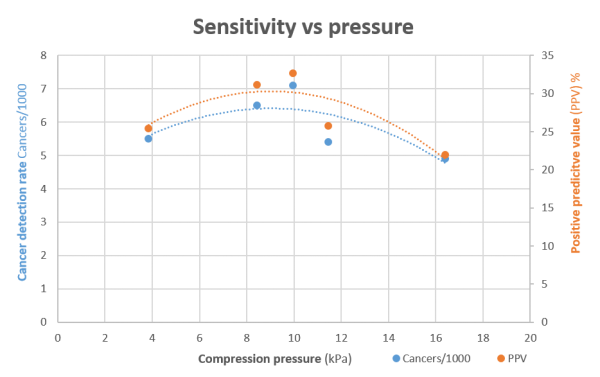
Figure 1: 10 kPa compression pressure during mammography corresponds to the peak optimum for cancer detection, in terms of cancer detection rate and positive predictive value.
The start of the second day saw Volpara Density featuring in two talks. Work at University of Manchester examined temporal change in volumetric density, concluding that “changes in mammographic density may be less important than initial mammographic density”. Karssemeijer’s group used Volpara to automatically detect textures within the breast that are independent of density. Following the discussions on density, Thomy Mertzanidou from University College London demonstrated a system to reconstruct 3D tumour structure post-operatively using histological slides derived from the lesion—a method that would greatly aid in improved ability to characterize cancers to better patient treatment. Other exciting technological developments (also from University College London) included a simulation that integrated MRI and 3D scans of the skin surface in order to predict the cosmetic outcome after breast conserving surgery. Data from four patients 6-12 months after surgery showed a mere 3.3 mm difference between the predicted and actual outcome (when using measurements from skin surface scans). There were also multiple in-depth discussions regarding contrast-enhanced and phase-contrast imaging, as well as the use of simulation studies for “virtual clinical trials”.
This meeting has been an excellent example of the ongoing innovation in the field of breast imaging and the role of new technologies. The demonstrated integration of knowledge from medical physics, computer science and machine learning, epidemiology and clinical science is a promising development in the fight against breast cancer.