Trying to assess breast density from a processed image is like a dermatologist trying to map a patient’s anatomy from a photograph that has been run through Instagram filters. The provider is seeing a nonquantitative processed depiction of patient anatomy that may not be explainable, repeatable, or true.
Volpara’s algorithms address this processing challenge by assessing the raw image. The use of raw images improves the accuracy of the quantification and reduces vendor variability to make it more generalizable, explainable, and repeatable.

There is no single standard for image processing. Mammograms are processed many different ways, and those methods are constantly evolving, vendor by vendor. The many types of technical design—anode, filter, geometry, kV, mAs, grid, detector—further complicate results.
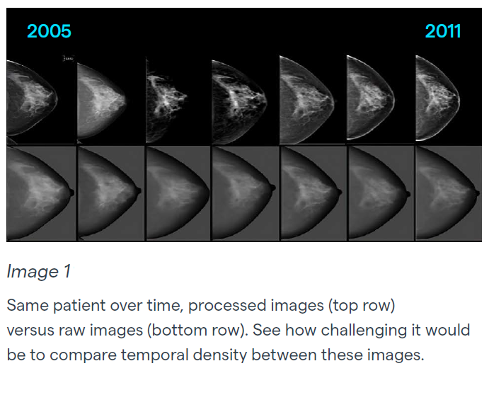
The impact of image processing can be seen most clearly when comparing images over time. Image 1 shows the same breast imaged over time, sometimes using different vendors. In the upper half of the graphic, the images have been processed and vary, sometimes significantly. In the lower half of the graphic, the images are all in their raw form and appear quite uniform.
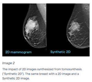
Image 2 shows the impact of comparing a 2D image to a synthetic 2D. Synthetic 2D images are not acquired images but are generated from the tomosynthesis image. Post processing is applied which can often suppress the visual appearance of density and may affect assessment.
I recently read a study about an AI algorithm being tested and the site seeing the number of cases being called suspicious doubling overnight. The culprit? A significant software update with a post processing change by the x-ray manufacturer unbeknownst to the AI vendor. And that is why the raw data is so, so important.
Discussions about how artificial intelligence (AI) can improve breast screening and breast radiology often stalls at the issue of vendor variability. RSNA’s Quantitative Imaging Biomarkers Alliance (QIBA), for example, stresses the need to understand all the potential imaging variations to get good AI. Similarly, scientists who only have images archived in PACS often use algorithms that recover what they can of the “preferred” raw images to derive quantitative data for breast density and texture patterns instead of what may have been introduced by vendor processing.
There’s no need to implement AI based on non-quantitative processed images that were developed to optimize viewing by a human. Ask us today about raw image acquisition and minimum network requirements.
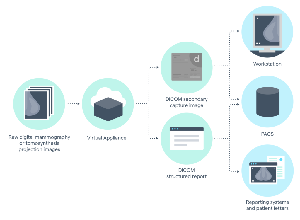
Data flow from raw images to secondary capture / report outputs